Snottite and Biovermiculation Microscopy and Spectroscopy
Click on the images to see the larger versions
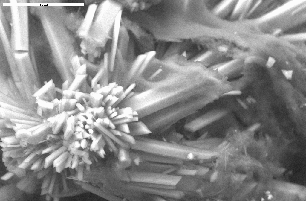
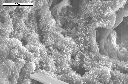
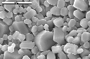
SEM image (15000x) of the internal structure of a snottite sampled in April,
1998. Note the gypsum crystals with the gauze-like substance woven in amongst
the crystals. We presume this gauze-like substance is a more refractory form
of polysaccharides. Photo by Spilde, Northup, and Boston. (973k)
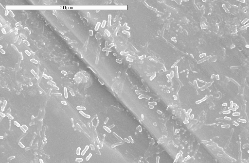
SEM image (10,000x) of a long view of a field of bacteria in the snottites.
Closeup images of a portion of this field are available below. Sampled
April, 1998 by Northup and Boston. Photo by Spilde, Northup, and Boston.
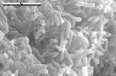
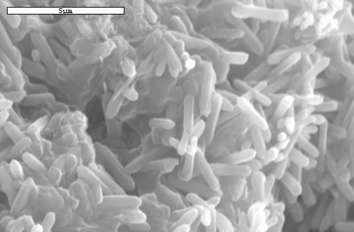
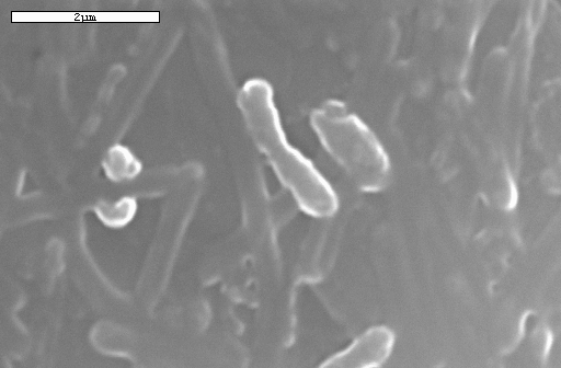
SEM image (13,000x) of bacterial bodies located in a snottite sampled
April, 1998 by Northup and Boston. Photo by Spilde, Northup, and Boston.

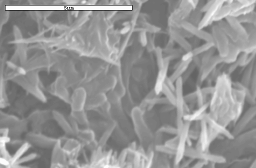
SEM image (15,000x) closeup of above bacterial bodies and mineral material.
Photo by Spilde, Northup, and Boston.
Putative bacteria on gypsum needles sampled January, 1998 near some snottites.
Photo by Spilde, Northup, and Boston.
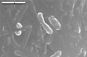
Enlargement of putative bacteria on gypsum needles sampled January, 1998
near some snottites.
Photo by Spilde, Northup, and Boston.
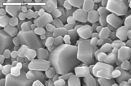
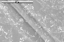
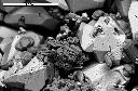
SEM image (5000x) of white gypsum paste sampled in January, 1998 next to the
snottites. The pH of this paste can be so low that it zeros out our field pH
meter. Photo by Spilde, Northup, and Boston.
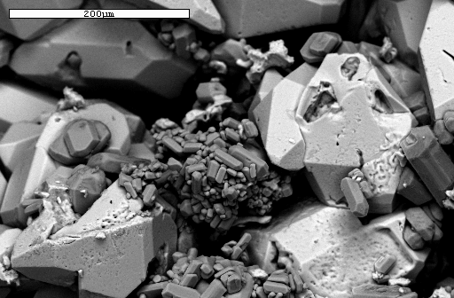
SEM backscattered electron image (300x) of sulphur (white) and gypsum
(medium gray) crystals from a sample (January 1998) of sulfur from the wall
of the cave. Photo by Spilde, Northup, and Boston.
Epifluorescent Images of Bacteria
in Biovermiculations
For questions or comments, please send mail to:
diana@i-pi.com